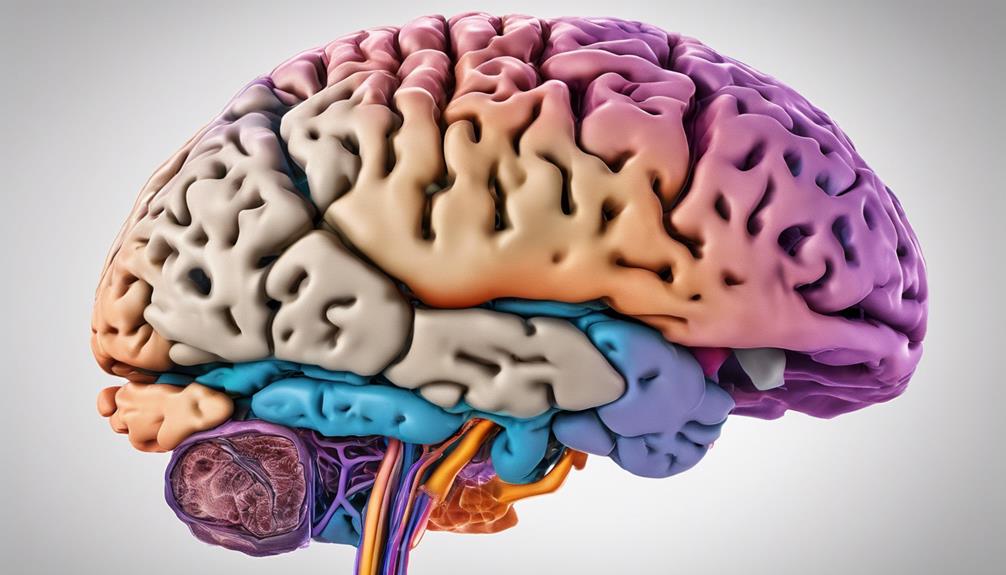

Exploring brain imaging to distinguish between normal brain function and dementia.
the intricate details revealed by these imaging techniques offer a fascinating glimpse into the complexities of the human brain.
The stark disparities observed between dementia-afflicted brains and those with normal cognitive function serve as a compelling starting point for a deeper exploration of how these scans can unravel the mysteries of neurological disorders.
The subtle nuances captured in these images hold the key to unraveling the intricate web of differences between a healthy brain and one grappling with the challenges of dementia.
Key Takeaways
- Brain atrophy indicates dementia progression.
- Enlarged ventricles signify tissue loss in dementia.
- Structural abnormalities disrupt neuronal communication in dementia.
- White matter lesions in dementia scans indicate brain damage.
Types of Brain Imaging Scans for Dementia
When diagnosing dementia, various brain imaging modalities, such as CT scans and MRIs, play a pivotal role in detecting structural abnormalities and changes indicative of the condition. CT scans utilize X-rays to capture detailed images of the brain, highlighting any structural irregularities associated with dementia.
Conversely, MRIs offer high-resolution images that aid in identifying brain atrophy, stroke damage, and other dementia-related alterations. Additionally, functional imaging techniques like PET scans, SPECT, and fMRI are instrumental in assessing brain function and blood flow, providing valuable insights into the physiological changes occurring in dementia patients.
These imaging modalities not only assist in the early detection of dementia but also contribute to accurate diagnosis and monitoring of disease progression by detecting biomarkers such as amyloid plaques. By utilizing a combination of these imaging tools, healthcare providers can offer comprehensive care to individuals with dementia, facilitating timely interventions and personalized treatment strategies.
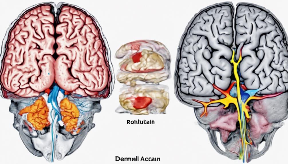
Key Differences in Dementia Brain Scans
In dementia brain scans, significant indicators such as brain atrophy, ventricle enlargement, and abnormal structures like plaques and tangles are commonly observed. These abnormalities provide crucial insights into the underlying changes associated with dementia.
- Brain Atrophy: Dementia brain scans often reveal a reduction in brain volume, particularly in regions responsible for memory and cognition. This shrinkage is indicative of neuronal loss and can impact overall brain function.
- Ventricle Enlargement: Enlarged ventricles, the fluid-filled spaces in the brain, are frequently seen in dementia scans. This enlargement reflects brain tissue loss and can affect the brain's ability to regulate cerebrospinal fluid, potentially leading to cognitive decline.
- Abnormal Structures: The presence of plaques and tangles in dementia brain scans is a hallmark of diseases like Alzheimer's. These abnormal protein deposits disrupt neuronal communication and contribute to cognitive and behavioral changes in patients.
Analyzing these key differences in dementia brain scans aids in diagnosing the condition early and devising appropriate treatment strategies to address both structural and functional abnormalities.
Importance of Early Dementia Detection
Pivoting from the discussion on key differences in dementia brain scans, detecting structural abnormalities and changes in brain function early through advanced imaging techniques is critical in optimizing interventions for individuals at risk for dementia.
Early detection of dementia is paramount as it allows for timely interventions and tailored management strategies. Brain scans play a pivotal role in differentiating between normal aging brain changes and pathological changes indicative of dementia.
Identifying dementia in its early stages is crucial for implementing effective treatment plans and enhancing the overall quality of life for patients. These scans unveil structural abnormalities and alterations in brain function that serve as early markers for dementia.
Progression Tracking Through Brain Scans
Utilizing advanced brain imaging techniques such as MRIs and CT scans allows for the precise tracking of structural changes and progressive brain atrophy over time in individuals with dementia. This monitoring is crucial for understanding the progression of the disease and evaluating the effectiveness of treatments.
- Detecting Alzheimer's Disease: Brain scans play a vital role in identifying the onset and progression of Alzheimer's disease through the observation of specific changes in the brain's structure.
- Assessing Disease Progression: Comparing current brain scans to previous ones enables healthcare providers to assess the rate of brain atrophy, providing valuable insights into the advancement of dementia.
- Monitoring Treatment Efficacy: By monitoring changes in the brain through scans, doctors can evaluate the impact of interventions, adjust treatment plans accordingly, and optimize patient care.
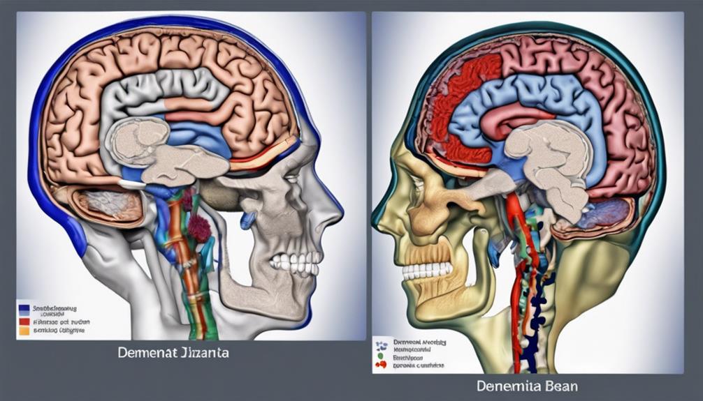
Understanding Normal Vs Dementia Brain Scans
Moving from the assessment of disease progression to the comparison of brain scans between normal individuals and those with dementia highlights distinct structural discrepancies indicative of the condition's impact on brain integrity. Normal brain scans typically exhibit a consistent brain structure devoid of significant abnormalities or signs of shrinkage.
In contrast, dementia brain scans often showcase brain atrophy, ventricular enlargement, and abnormal changes in specific brain regions. These deviations can manifest as altered brain volume, reduced cortical thickness, and compromised overall structural integrity. Additionally, dementia brain scans may reveal visible signs of damage such as white matter lesions not commonly observed in normal scans.
Understanding these disparities between normal and dementia brain scans is crucial for accurate dementia diagnosis and effective treatment planning. By analyzing the differences in brain structure and integrity, healthcare professionals can better tailor interventions to address the specific needs of individuals affected by dementia, ultimately enhancing their quality of life.
Frequently Asked Questions
Can You Tell by a Brain Scan if You Have Dementia?
Yes, brain scans can reveal signs of dementia by detecting brain shrinkage, atrophy, or abnormal structures. Specific patterns on scans indicate various types of dementia, like Alzheimer's or vascular dementia.
Amyloid PET scans identify amyloid plaques, a hallmark of Alzheimer's. Changes in brain structure visible on scans aid in early detection before symptoms appear.
Scans are vital for ruling out other conditions and tracking dementia progression.
How Your Body Warns You That Dementia Is Forming?
As our bodies undergo the early stages of dementia formation, subtle signs may emerge. These warning signals can manifest as changes in memory, language, behavior, and decision-making abilities.
Mood swings, disorientation, and withdrawal from social activities may also indicate the onset of dementia. Recognizing these indicators and seeking timely medical intervention can aid in the early detection and management of this condition.
What Do Dementia Eyes Look Like?
Dementia eyes typically exhibit reduced blinking, lack of focus, and a distant or vacant appearance. Changes in eye movements, like slow or uncoordinated gaze shifts, may also be noticeable. These manifestations reflect cognitive decline and difficulty processing visual information.
Individuals with dementia might struggle to make eye contact or engage with their surroundings. Understanding these eye-related signs can aid in recognizing and addressing cognitive impairments associated with dementia.
What Is the First Noticeable Symptom of Dementia?
Memory loss, akin to a scattered jigsaw puzzle, is often the initial sign of dementia. This puzzle, when incomplete, hints at the complexities within the brain.
Understanding this crucial clue allows for early interventions, enhancing the quality of life for those affected. Recognizing this symbol of cognitive decline prompts us to engage in proactive measures, seeking medical guidance to navigate the intricate landscape of aging and neurological health.
Conclusion
In conclusion, the intricate dance of neurons captured within brain scans serves as a symphony of clues in the diagnosis of dementia. Like skilled conductors, these imaging techniques reveal the discordant notes of structural changes, guiding clinicians towards tailored treatment plans.
As we unravel the enigmatic composition of the brain, we gain a deeper appreciation for the symmetrical beauty of normal brain function and the haunting melody of dementia's progression.

Ethel leads our content team with a keen eye for detail and a passion for storytelling. With years of experience in editing and content development, Ethel ensures that our resources are informative, engaging, and accessible to caregivers from all walks of life.